
Why Your Family Dentist Uses Digital X-Rays for Safer Checkups

Why Dentists Use Digital X-Rays for Family Checkups
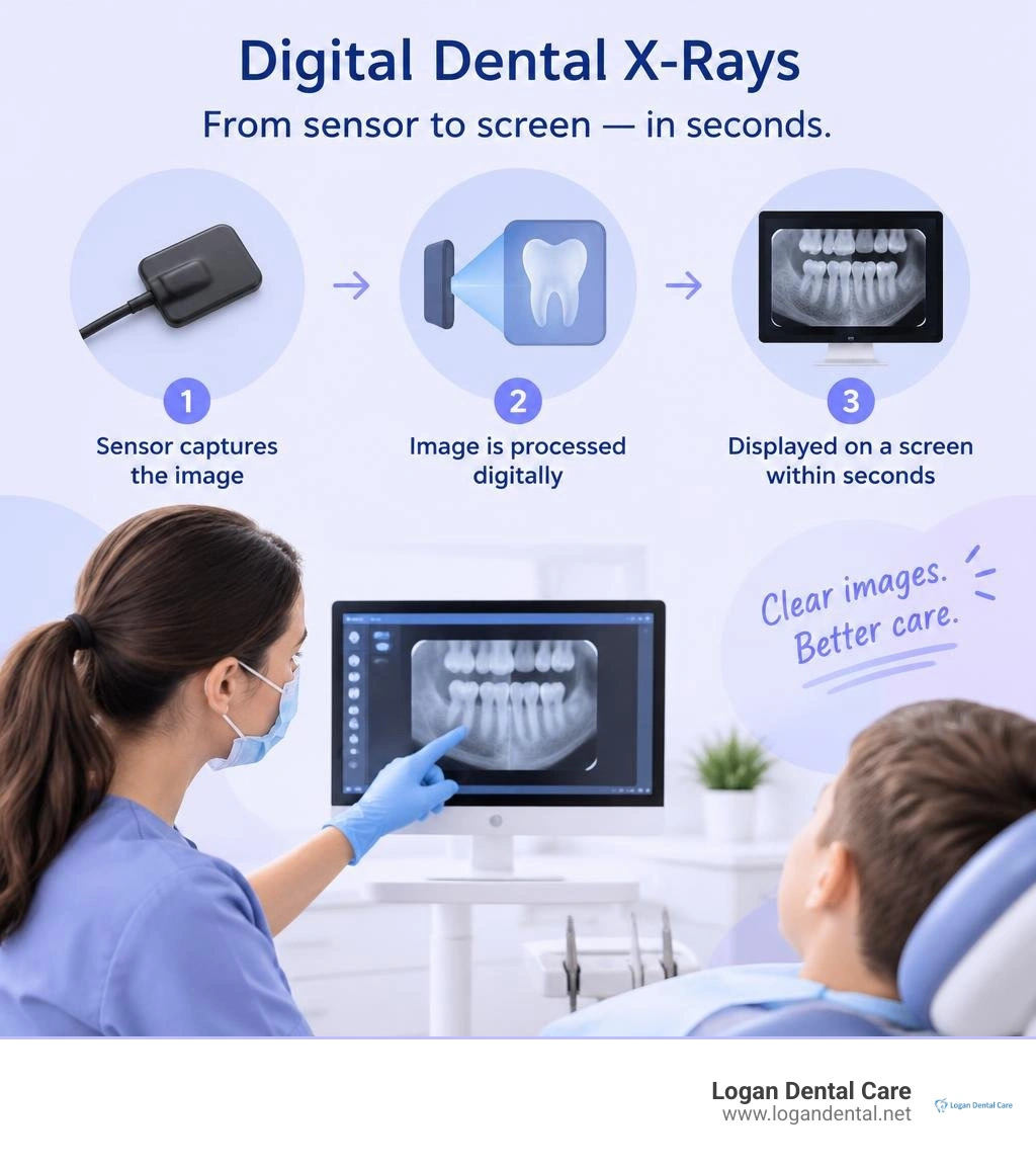
Digital X-rays at the dentist use an electronic sensor instead of film to show images of teeth and jawbone on a screen within seconds. They can reveal cavities between teeth, infections, and bone loss that a visual exam may miss. Digital imaging generally uses less radiation than film and needs no chemical developing. Your dentist should still recommend X-rays only when they are needed for your care.
Logan Dental Care is a family-focused practice at 1021 N. Main Street in Bellefontaine, Ohio, serving Bellefontaine and surrounding Logan County communities. The practice is an accredited member of the American Dental Association and the Ohio Dental Association and holds an A+ BBB rating. We're the Logan Dental Care Team, and we'll walk you through what digital imaging means for your family's checkups.
Learn more about digital x rays dentist:
Understanding Digital Radiography vs. Traditional Film Imaging
Dental imaging has evolved significantly since the first commercial digital dental imaging system was introduced in 1987. In the past, dentists placed small packets of film inside your mouth, exposed them to an X-ray beam, and developed the film in a darkroom using chemical processing tanks. This process took several minutes and produced physical plastic sheets that had to be filed away in paper folders.
Modern dental practices rely on solid-state digital sensors, such as charge-coupled devices (CCD) or complementary metal-oxide-semiconductor (CMOS) technology, as well as photostimulable phosphor storage plates. When you visit a practice that uses our technology, the digital sensor captures X-ray photons and immediately sends the data to a chairside computer screen. This direct conversion provides crystal-clear images in seconds, completely eliminating the need for toxic chemical fixers and developers (Jayachandran, 2017). For a broader look at how radiography has progressed, you can read the Digital Imaging in Dentistry Review.
Key Differences Between Digital Sensors and Traditional Film
Digital radiography offers distinct clinical advantages over conventional film packets:
- Speed of Image Capture: Digital sensors capture and render images on a display screen almost instantly, reducing your time in the dental chair.
- Dynamic Range and Image Adjustments: Digital detectors possess a dynamic range up to 400 times wider than traditional film-screen systems. This allows your dentist to adjust contrast, brightness, and sharpness without taking another exposure.
- Magnification Tools: Dentists can zoom in on fine anatomical structures, such as subtle root fractures or early enamel demineralization, with exceptional clarity.
- Environmental Safety: Digital imaging eliminates lead foil wrappers, plastic packets, and liquid chemical waste, making it much more environmentally friendly.
Why Choosing a Digital X-Rays Dentist Ensures Safer Care
Patient safety is at the heart of modern dentistry. One of the greatest benefits of digital imaging is the dramatic reduction in radiation exposure. Digital X-rays reduce radiation by 80% to 90% compared to traditional film radiography. A conventional dental film exposure emits about 0.5 millirem (mrem), whereas a digital sensor requires as little as 0.1 mrem.
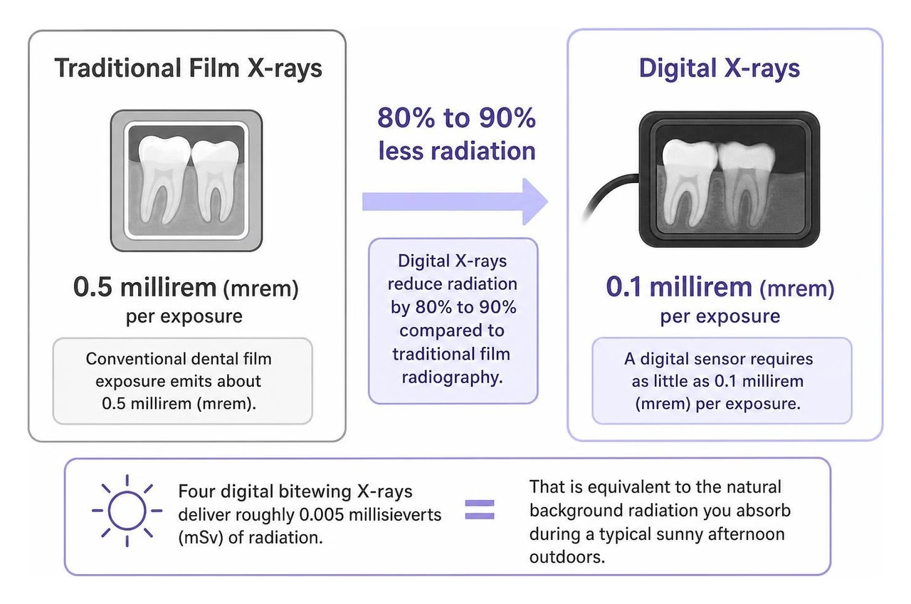
To put this in perspective, four digital bitewing X-rays deliver roughly 0.005 millisieverts (mSv) of radiation. That is equivalent to the natural background radiation you absorb during a typical sunny afternoon outdoors. Dental imaging accounts for less than 1% of the annual medical radiation dose received by individuals in the United States.
Dentists follow the ALARA principle—keeping exposure "As Low as Reasonably Achievable." By combining high-sensitivity sensors with rectangular collimation, which limits the beam size strictly to the sensor area, practices can reduce radiation scatter by more than 60%. Guidelines published in the ADA / FDA Guide to Patient Selection for Dental Radiographs | FDA help ensure exposures occur only when clinically justified.
Safety Standards for Children and Expecting Mothers
Protecting growing children and expectant mothers requires thoughtful care and strict safety protocols. Pediatric dentistry follows the national "Image Gently" initiative, which sizes radiation doses specifically for smaller bodies and developing jawbones. Our family dentistry approach focuses on child-sized sensor positioning and personalized imaging intervals tailored to each child's individual cavity risk.
For pregnant patients, routine screening radiographs are often postponed until after delivery. However, if a dental emergency or infection arises, diagnostic digital X-rays can be safely taken. Because the digital sensor requires minimal exposure and protective shielding prevents scatter, dental diagnostics remain safe when treating urgent conditions during pregnancy.
Intraoral vs. Extraoral Imaging: Types of Modern Dental X-Rays
Dentists use two primary categories of radiographs: intraoral (where the sensor rests inside the mouth) and extraoral (where the imaging device rotates around the outside of your head). To learn more about various imaging modalities, review the Dental X-rays: Purpose & Types guide from the Cleveland Clinic.
Intraoral Radiographs for Tooth and Bone Health
Intraoral views provide close-up diagnostic detail for individual teeth and surrounding bone:
- Bitewing Radiographs: These images capture the crowns of your upper and lower teeth together. They are essential for detecting cavities forming between touching teeth and tracking the height of supporting bone during routine dental hygiene visits.
- Periapical (PA) Radiographs: These show the entire tooth from crown to the tip of the root where it anchors into the jaw. They help diagnose root abscesses, internal nerve damage, and deep bone cysts.
- Occlusal Radiographs: These broader intraoral images show the full floor or roof of the mouth, helping dentists track unerupted teeth or developmental irregularities in children.
Extraoral Imaging and 3D Cone-Beam CT (CBCT)
Extraoral imaging captures broad anatomical views of your jaw, skull, and facial structures:
- Panoramic X-Rays: A machine rotates around your head to create a single broad overview of your upper and lower jaws, sinuses, and temporomandibular joints (TMJ). They are ideal for evaluating wisdom teeth, jaw alignment, and bone pathology.
- Cephalometric Projections: These side-profile images capture the relationship between teeth and facial bones, aiding orthodontists in planning corrective treatments.
- Cone-Beam Computed Tomography (CBCT): CBCT provides three-dimensional, high-resolution scans of teeth, nerve pathways, and bone volume. Dentists use 3D CBCT scans for precise dental implant planning, complex root canal therapy, and reconstructive surgery.
How Digital Dental Imaging Transforms Diagnosis and Treatment Planning
Digital radiography connects seamlessly with dental software. Because digital images use the standardized DICOM format, they integrate directly into electronic patient records. This enables dentists to track bone levels, compare subtle enamel changes side-by-side over time, and immediately share files with oral surgeons or orthodontists when coordinating specialized care.

How a Digital X-Rays Dentist Detects Hidden Oral Health Issues Early
A visual exam only reveals what is happening on the outer surfaces of your enamel. Digital imaging allows your dentist to see deep inside and between teeth to uncover:
- Early interproximal decay between tight contacts before pain begins
- Hidden infections or bone loss around roots, allowing for prompt dental plaque treatments
- Recurrent decay forming beneath aging crowns or fillings
- Impacted teeth resting below the gumline
- Bone changes associated with periodontal disease
Spotting these conditions early allows for conservative, gentle treatments rather than extensive dental procedures later on.
What to Expect When Visiting a Digital X-Rays Dentist for Routine Exams
Getting digital X-rays is fast, simple, and comfortable. During your preventive appointment, a dental assistant or hygienist covers the slim digital sensor with a soft, protective hygienic sleeve. The sensor is placed next to your teeth while you bite down gently on a comfortable holder.
The machine activates for a fraction of a second, and the image immediately appears on the chairside monitor. If you have a sensitive gag reflex, breathing slowly through your nose and working with a patient, caring team makes the experience quick and easy as part of your daily dental care integrating oral health into your self-care routine.
Frequently Asked Questions About Digital Dental X-Rays
How often do I really need to have dental X-rays taken?
The American Dental Association recommends custom imaging schedules based on individual oral health. Healthy adults with a low risk of tooth decay typically need bitewing X-rays only every 24 to 36 months. Patients with active periodontal disease, frequent new cavities, or high risk factors may benefit from imaging every 6 to 18 months to catch issues early. Your dentist will provide tailored oral health advice based on your unique smile.
Can I refuse dental X-rays during my checkup?
Yes, as a patient, you always have the right to refuse dental X-rays. However, dentists rely on X-rays to see bone levels, tooth roots, and interproximal surfaces that cannot be evaluated with the naked eye. Refusing necessary diagnostic imaging limits your dentist's ability to diagnose hidden disease and may prevent them from providing safe, comprehensive restorative care that meets professional standards.
Do digital dental X-rays cost more than conventional film X-rays?
No. Digital dental X-rays are billed under standard diagnostic codes, meaning out-of-pocket costs are equivalent to or lower than older film methods. Most dental benefit plans cover preventive diagnostic radiographs at 100% during routine cleanings. For more context on common preventive care fees, review our dental cleaning cost guide 2026.
Conclusion
Digital X-ray technology provides a safer, faster, and more comfortable diagnostic experience for patients of all ages. By reducing radiation exposure up to 90%, eliminating harsh darkroom chemicals, and delivering crystal-clear images in seconds, digital imaging makes modern preventive dental care more precise and collaborative.
Logan Dental Care is a family-focused dental practice located at 1021 N. Main Street in Bellefontaine, Ohio, serving Bellefontaine and surrounding Logan County communities. Logan Dental Care is an accredited member of the American Dental Association and the Ohio Dental Association, and holds an A+ BBB rating.
Our team provides comprehensive general and preventive dentistry, family dentistry, cosmetic dentistry (teeth whitening, veneers, smile makeovers), orthodontics including traditional braces and Invisalign clear aligners, restorative dentistry (implants, crowns, bridges), emergency dental care with same-day and after-hours availability, nightguards and mouthguards, and the Smile Protection Savings Plan for uninsured patients. Please note that we do not offer payment plans or sedation dentistry.
If it has been a while since your family's last dental checkup, contact us today to schedule your next visit and experience warm, modern, and gentle dental care.
References
Jayachandran S. "Digital Imaging in Dentistry: A Review.." Contemporary clinical dentistry, 2017. PMCID PMC5551320.